Medical devices
search
news
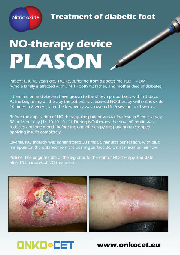
The PDF with the short report with pictures from the therapy of a diabetic foot can be viewed or downloaded here.
The pictures from the treatment of unhealing wounds an be found here:
http://www.onkocet.eu/en/produkty-detail/220/1/
The pictures from the treatment of unhealing wounds an be found here:
http://www.onkocet.eu/en/produkty-detail/293/1/

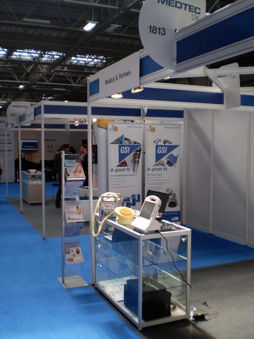
ONKOCET Ltd. has exhibited the devices from its portfolio on the MEDTEC UK exhibition in Birmingham, April 2011 through our partner Medical & Partners.
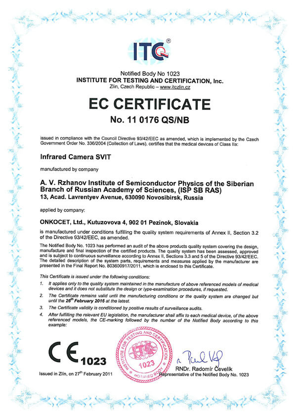 The ONKOCET company has successfully reached the certification of yet another medical device, Infrared Camera SVIT. The Certificate can be found here. The videos from the device operation can be found here.
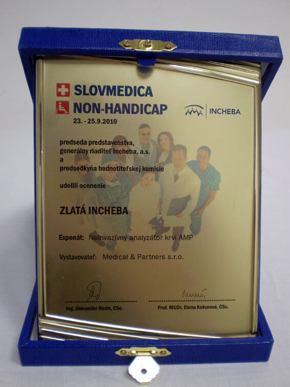 Our device, the non-invasive blood analyzer AMP has won the Golden Incheba prize at a medical exhibition SLOVMEDICA - NON-HANDICAP 2010. A big thank you goes to the organizers of the exhibition for acknowledging the quality of our device and to the exhibitor, the Medical & Partners company, for introduction of the AMP device to the medical public again.
We are pleased to inform our business partners, that our company has succesfully finished the certification process of Concor Soft Contact Lenses.
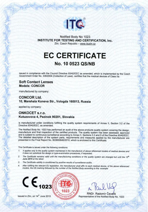 You can find the certificate here.
More information on Concor Soft Contact Lenses go to section Medical preparations/Concor soft contact lenses, or follow this link.
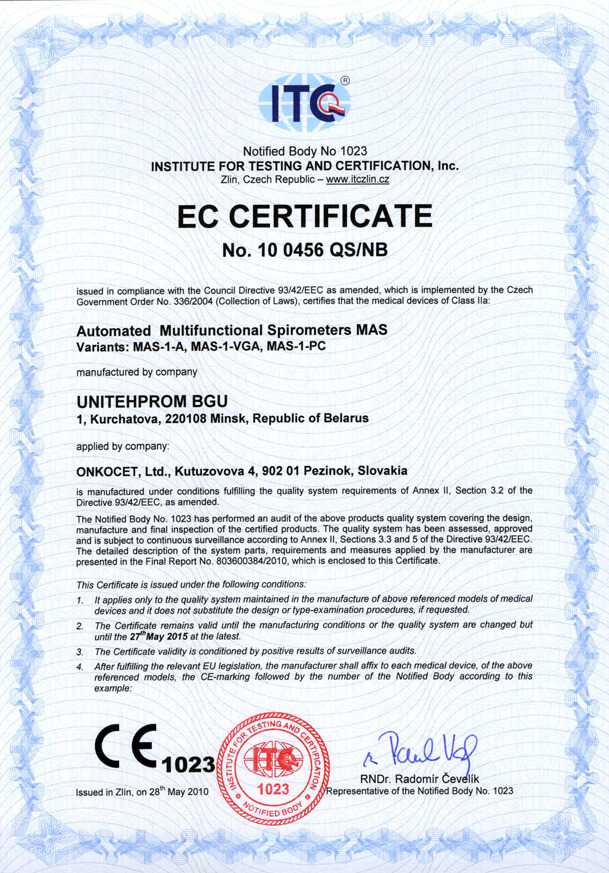 Our company has finished the certification process for another medical device, computerized spirometer MAS-1K with oximeter. You can find the device certificate here.
.jpg) Since May 2010 there is a new version of AMP device available.
Since May 2010 there is a new version of AMP device available.Follow this link if you want to see the pictures and specifications of the device.
http://www.onkocet.eu/en/produkty-detail/293/1/
 Dear partners,
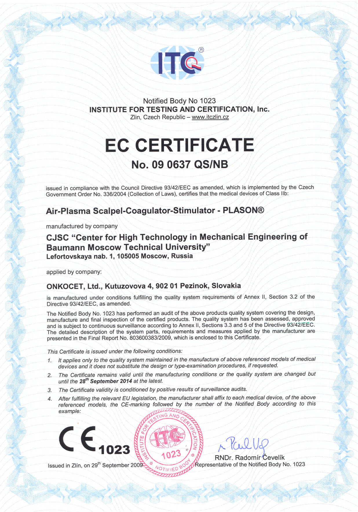
 In October 2009 we have received CE certificate for another device from our portfolio, NO therapeutical device PLASON. You can find more information about this revolutionary device, used for healing of unhealing wounds, diabetic foot, or for cosmetical purposes, at our webpage, section "Medical devices" -> PLASON-NO Therapy.
.gif)
Best regards
Team of ONKOCET Ltd. company
Other
TECHNICAL SUMMARY DIGEST
Biomedical Optics
19-25 January 2002 San Jose Convention Center San Jose, California USA
Symposium Chairs: Warren S. Grundfest., Cedars-Sinai Medical CenterJoseph R. Lakowicz, Univ. of Maryland School of Medicine / Baltimore
SPIE's BiOS 2002
4618-08, Session 3
 The goal of this work is to analyze the capabilities of a radiometric system for remote temperature control during laser thermal therapy. The main feature of this system is the combination of several spectral channels: near IR, middle IR and microwave range. The first and second channels provide information about the temperature distribution around the surface and subsurface of absorbing structures like hair follicles and pigmented lesions. The third channel provides information about the localized laser-induced temperature effects occurring much deeper, up to 3-5 cm. If the area of interest lies deep within the tissue the first and second channels are used for calibration purposes only. Experimental data in vitro using a near IR diode laser are presented, focusing on the estimated capabilities of the new radiometric monitor as a system to provide feedback control for enhanced interstitial laser therapy of local tumors. Further modifications of this system are suggested for photothermal (PT) radiometric confocal microscopy. The modifications is based upon the combination of PT radiometry in time-resolved, and frequency-domain modes and confocal microscopy using reflected scanning modes. The potential advantage of these new approaches are discussed including the imaging of tissue chromophores with high spatial resolution based upon measurements of the thermal gradients created by localized absorption at varying depths...
Photothermal/Microwave Radiometry for Imaging and Temperature Control
PDF file: 630 kB![]()
First In Vivo Application of Microwave Radiometry in Human Carotids
A New Noninvasive Method for Detection of Local Inflammatory Activation
Konstantinos Toutouzas, MD,* Charalampos Grassos, MD,* Maria Drakopoulou, MD,* Andreas Synetos, MD,* Eleftherios Tsiamis, MD,* Constantina Aggeli, MD,* Konstantinos Stathogiannis, MD,* Dimitrios Klettas, MD,* Nikolaos Kavantzas, MD,†Georgios Agrogiannis, MD,† Efstratios Patsouris, MD,† Christos Klonaris, MD,‡ Nikolaos Liasis, MD,* Dimitrios Tousoulis, MD,* Elias Siores, BSC, MSC, PHD,§ Christodoulos Stefanadis, MD*
Athens, Greece; and Bolton, United Kingdom
 Objectives
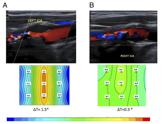
This study investigated whether temperature differences: 1) can be measured in vivo noninvasively by microwave radiometry (MR); and 2) are associated with ultrasound and histological findings.
Background
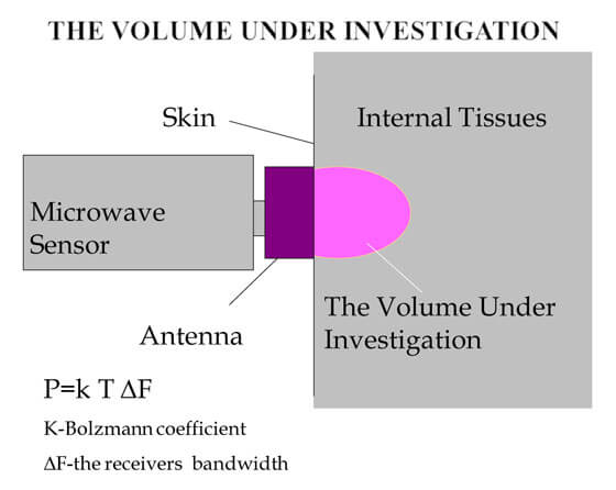
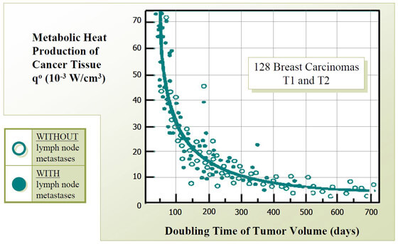
Studies of human carotid artery samples showed increased heat production. MR allows in vivo noninvasive measurement of internal temperature of tissues.
Methods
Thirty-four patients undergoing carotid endarterectomy underwent screening of carotid atherosclerosis by ultrasound and MR. Healthy volunteers were enrolled as a control group. During ultrasound study, plaque texture,plaque surface, and plaque echogenicity were analyzed. Temperature difference was assigned as maximal minus minimum temperature. Association of thermographic with ultrasound and histological findings was performed.
Photothermal/Microwave Radiometry for Imaging and Temperature Control
PDF file: 1 MB
![]()
Microwave radiometry: a new non-invasive method for the detection of vulnerable plaque
Konstantinos Toutouzas, Andreas Synetos, Charalampia Nikolaou, Konstantinos Stathogiannis, Eleftherios Tsiamis, Christodoulos Stefanadis
First Department of Cardiology, University of Athens, Medical School, Hippokration Hospital, Athens, Greece
Corresponding to: Konstantinos Toutouzas, MD. 26 Karaoli and Dimitriou str., Holargos, 15562, Athens, Greece. Email: ktoutouz@otenet.gr.
 Abstract: Atherosclerosis and its consequences are the most rapidly growing vascular pathology, with myocardial infarction and ischemic cerebrovascular accident to remain a major cause of premature morbidity and death. In order to detect the morphological and functional characteristics of the vulnerable plaques, new imaging modalities have been developed. Intravascular thermography (IVT) is an invasive method, which provides information on the identification of the high-risk atheromatic plaques in coronary arteries. However, the invasive character of IVT excludes the method from primary prevention. Microwave radiometry (MR) is a new non-invasive method, which detects with high accuracy relative changes of temperature in human tissues whereas this thermal heterogeneity is indicative of inflammatory atherosclerotic plaque. Both experimental and clinical studies have proved the effectiveness of MR in detecting vulnerable plaque whereas recent studies have also revealed its association with plaque neoangiogenesis as assessed by contrast enhanced carotid ultrasound (CEUS).
Key Words: Microwave radiometry (MR); intravascular thermography (IVT), neoangiogenesis; vulnerable plaque.
Source:
Toutouzas K, Synetos A, Nikolaou C, Stathogiannis K, Tsiamis E, Stefanadis C. Microwave radiometry: a new non-invasive method for the detection of vulnerable plaque. Cardiovasc Diagn Ther 2012;2(4):290-297. DOI: 10.3978/j.issn.2223-3652.2012.10.09
© Cardiovascular Diagnosis and Therapy.
to view the article follow this link
![]()
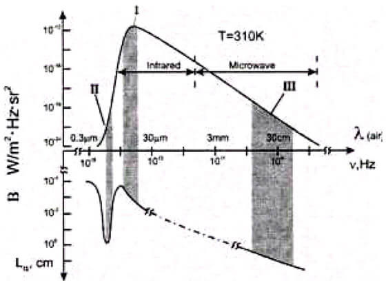
Basis of Microwave Radiometry
 Introduction
Our concept about RTM-Diagnosis is first of all based on our own investigations, which we have conducted over 6 years in five leading Moscow Oncological Centres. Over this period we have performed
3.500 RTM-examinations, and 540 women were cancer patients. Also our views were formed by investigations of Michel Gautherie, a French Scientist in Breast Thermobiology. He worked in this field for over 16 years. His data published in [1] is based on investigation of 85.000 patients. In 247 patients he measured the temperature invasively with the help of fine-needle thermoelectric probes. This unique set experimental data form the basis of our modern concepts about temperature distribution in the breast.
RTM Basis_of_Microwave_Radiometry.pdf
PDF File: 1.0 MB ![]()